What you’ll take away
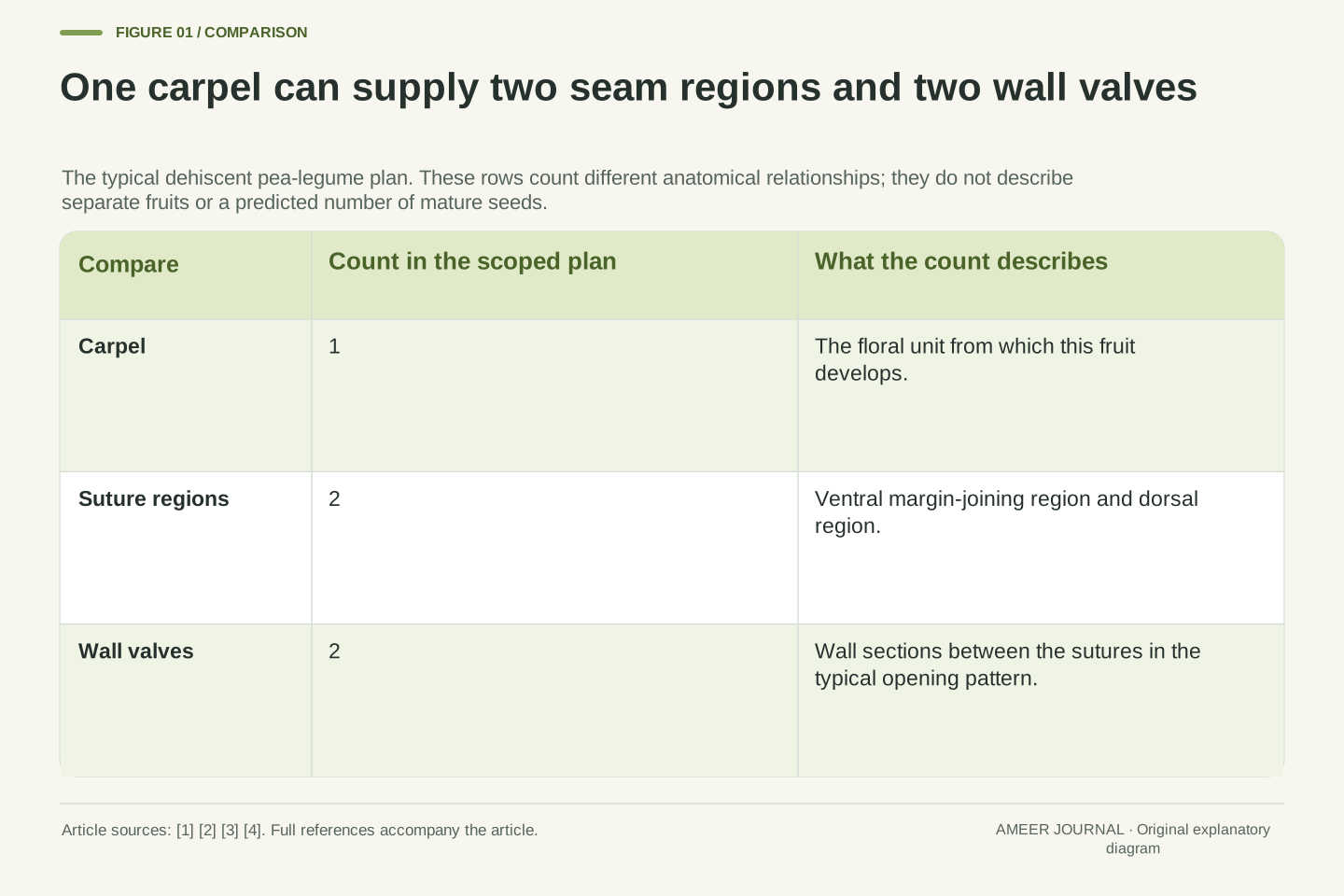
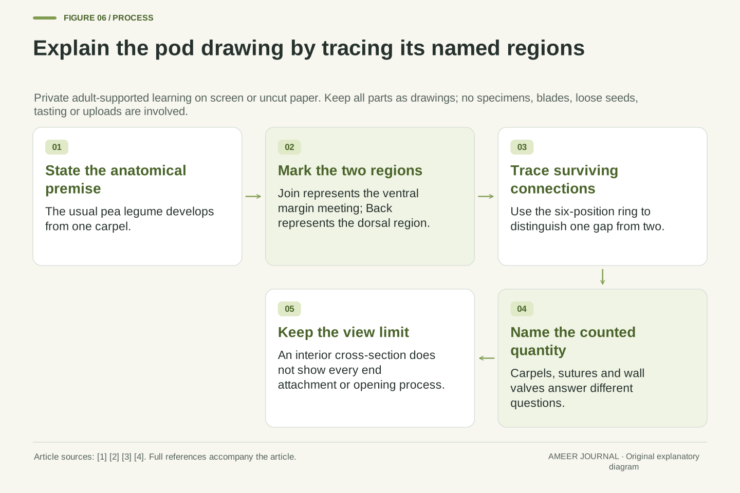
- The typical pea legume develops from one carpel even though it can open along two suture regions.
- The ventral region joins the carpel margins; the dorsal region has a different structural role.
- Two wall valves are not evidence of two original carpels.
- In the original ring, one gap leaves one connected path; two named gaps leave two arcs.
- A cross-section omits end attachments and does not predict a real fruit’s opening time.
Two opening lines do not automatically mean two starting parts
A pea pod has two long seam regions. In the usual opening legume pattern, its wall can separate along both of them. That might suggest the pod was made from two matching flower parts. Botanists describe something different: this fruit develops from one carpel, a seed-bearing structure of the flower [1][2]. The opening lines and the starting structure count different things.
Think about a drawn loop. It begins as one connected outline. If we leave gaps in two places, the remaining outline has two separate arcs. Those arcs did not need to begin as two different loops. This little drawing will help us explore the pod's wall, while the botanical sources tell us which parts and seam regions the drawing represents.
Read with a grown-up or another suitable learning partner. Use the illustrations on screen or make your own on an uncut sheet of paper. Keep every part as a drawing: no pods, loose seeds, plants, needles, scissors or tasting are needed. Your page can stay private at home; there is nothing to photograph, upload or register.
A typical dehiscent pea legume is monocarpellate: its fruit develops from a single carpel, while dehiscence follows two longitudinal sutures [1][2]. The sutures describe opening regions; the carpel describes a floral structural unit. Equating two sutures or two wall valves with two original carpels confuses different anatomical relationships.
This adult-supported lesson connects those definitions to an original boundary model. A labelled strip illustrates joined margins and a dorsal region; a six-position transverse ring then distinguishes closedness from connected wall portions. The geometry illustrates how the stated organization is possible. It does not independently establish the developmental identity of a plant specimen.
The activity is entirely on screen or uncut paper, with a learning partner. Numbered marks represent wall positions, not seeds, cells or children. No physical specimen is gathered or opened, no loose seeds or cutting tools are used, and no personal details or online submission are requested. The Technical mode adds anatomical terminology and explicit model limits.
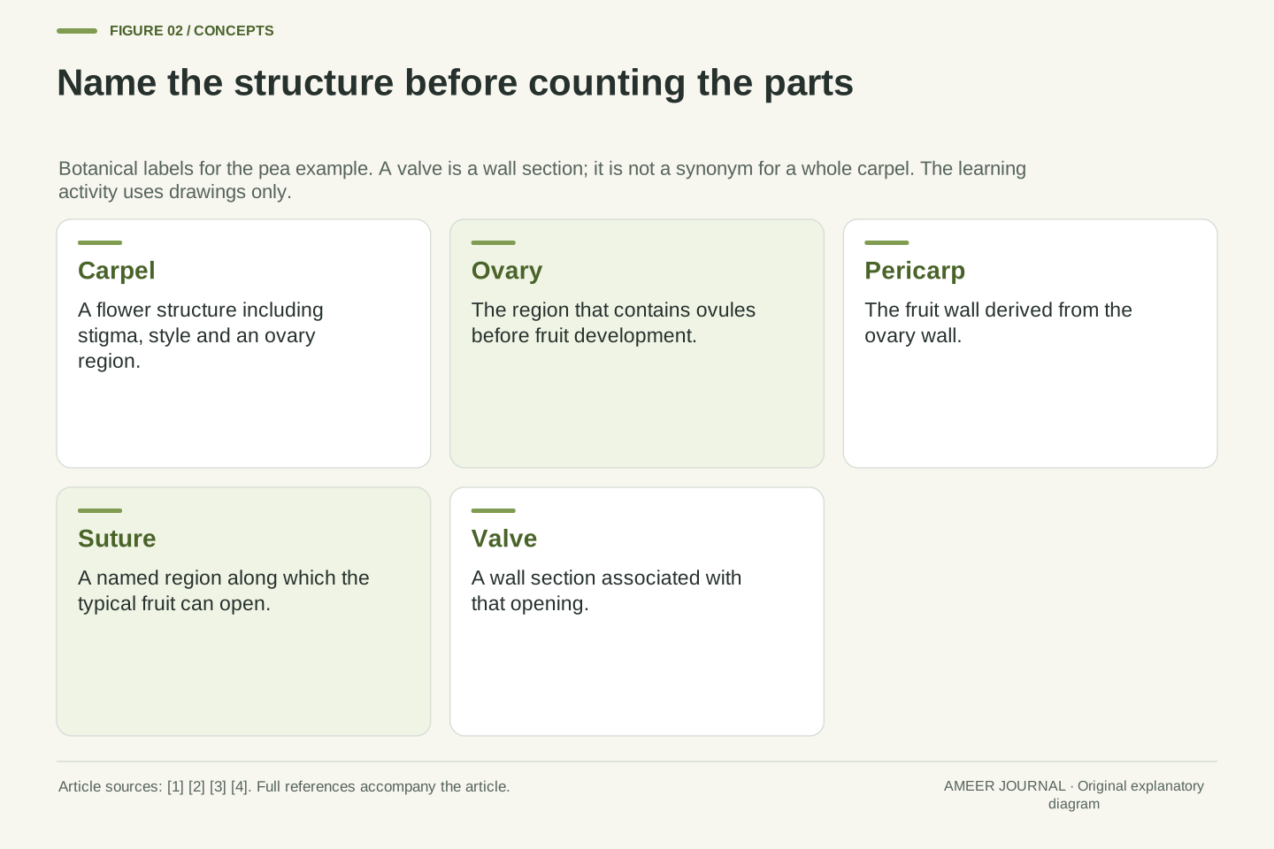
A carpel is a flower structure, not one half of an opened pod
A carpel includes the stigma, style and ovary. The stigma receives pollen; the style leads toward the ovary, which contains ovules [1][3]. For this question, focus on the containing structure. In the usual pea arrangement, one carpel provides the ovary whose wall later becomes the pod wall. Its two sides do not need to be two separate carpels.
Botanists call a mature fruit wall the pericarp [1]. That is a name for tissue, not a count of opening pieces. When the wall separates into sections, the sections are still portions of that fruit wall. Giving them a new descriptive name does not change the number of carpels from which this fruit developed.
There is also a wider naming trap. A fruit called simple can arise from one carpel or from several joined carpels [1]. Simple therefore does not always mean exactly one carpel. Our one-carpel statement belongs to the pea legume being explained, rather than to every fruit that looks like one object.
Carpel, ovary and pericarp are related but noninterchangeable terms. A carpel includes stigma, style and the ovary region; the ovary contains ovules. The fruit wall develops from the ovary wall and is termed pericarp [1][3]. A valve, introduced below, is a portion of that wall associated with opening, not a synonym for a whole carpel.
The source description calls the legume monocarpellate [2]. By contrast, a compound pistil can contain fused carpels, and the broader category simple fruit can include a single ovary formed by several fused carpels [1][3]. An external single-object count is therefore insufficient for a general carpel count.
Here we accept the source-supported single-carpel pea plan as the anatomical premise, then examine its consequences for seam interpretation. The drawing is not a method of discovering hidden floral anatomy from shape alone. Keeping the premise explicit prevents a topological example from being mistaken for a universal identification key.
Two margins can meet to make one seam
Imagine one long strip drawn flat on the page. Mark its two long edges Margin One and Margin Two. Also draw a line lengthwise through its middle. The strip is one represented sheet with two edges. We will imagine its sides curving toward one another until the two margins meet; the paper itself stays flat.
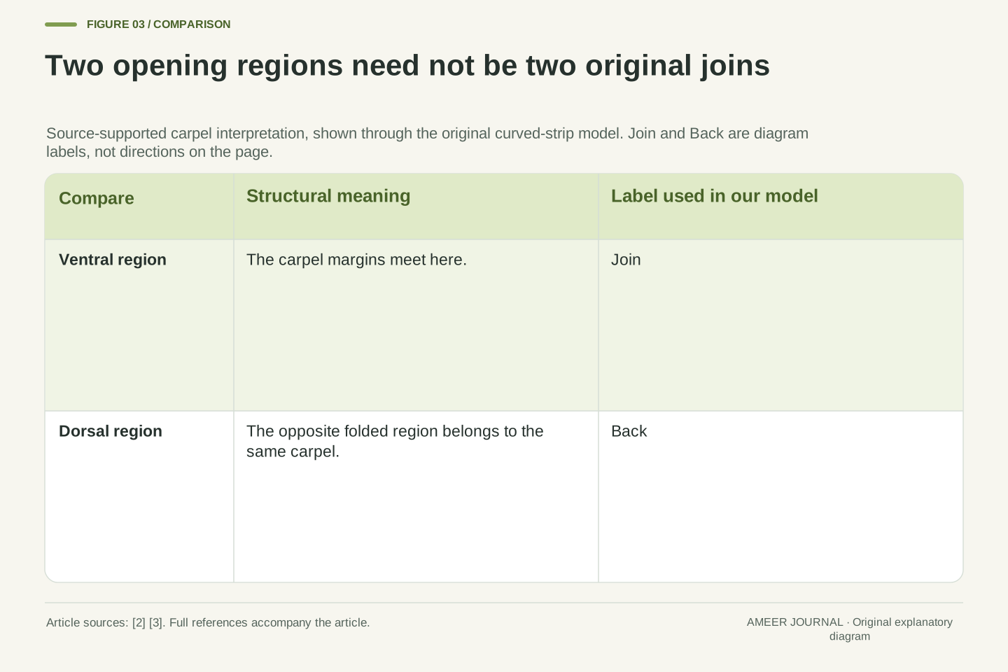
Where the margins meet, they form one joining line. Two edges have become the two sides of a single join, rather than two separate joins. In the botanical folded-carpel model, that joining region is the ventral suture [2][3]. We will give it the friendly label Join on our original drawing.
The middle line ends up on the opposite side of the curved wall. It does not vanish when the edges meet. Our sketch therefore contains a joining region and a separate back region while still representing one original sheet. This is a structural way to understand the carpel description, not a film of a living flower literally being folded by hand.
Parameterize the width of an original flat schematic strip from x=0 to x=8, in arbitrary drawing units. Identify the two longitudinal margins x=0 and x=8 along one seam labelled Join. The central line x=4 remains a distinct longitudinal region. The identification changes the boundary relationship without introducing a second original sheet.
The botanical interpretation assigns the fused-margin region to the ventral suture and the opposite folded region to the dorsal suture [2][3]. The source's folded-carpel description is used as an anatomical model. It is not asserted to be a measured developmental animation or a literal leaf-folding procedure performed by the learner.
Two pre-join margins, one margin-joining seam and one additional dorsal region are three different counts. In the schematic, the wall intervals 0≤x≤4 and 4≤x≤8 lie between the two named regions after identification. Equal widths follow our chosen drawing coordinates; they are not measured dimensions or a universal equality of real pod-wall areas.
The ventral and dorsal regions have different meanings
Join has a botanical name: ventral. The opposite back region is called dorsal. The university fruit lab describes ventral as the region where carpel margins fuse and dorsal as the region of folding [2]. They can both be opening lines without having the same structural origin.
This is an important detail about the word seam. In this lesson it can name a line along which a fruit opens. It does not always mean that two originally separate pieces were glued together there. The dorsal line belongs within the same represented carpel wall; it is not a second margin join created by bringing in another carpel.
Add two different labels to the curved-strip drawing: Join for the ventral region and Back for the dorsal region. Use words or different line styles so the drawing works without relying on colour. Ask your partner to point to the line where the margins meet, then to the other line. Explaining that difference answers more than merely counting two lines.
For the scoped legume morphology, ventral refers to the region of carpellary margin fusion, while dorsal refers to the opposing folded region [2][3]. The two longitudinal dehiscence sutures can therefore occur within a fruit derived from one carpel. The existence of a dorsal opening does not establish fusion between two originally distinct carpels.
Keep the descriptive and developmental uses of a line separate. A line of opening is a location where the mature wall separates; a margin-fusion line describes an anatomical relationship in the carpel model. The ventral region plays both roles in the typical example. The dorsal region supplies the second opening route without becoming a second fused-margin boundary.
Our labels Join and Back preserve those identities throughout every later sketch. Their placement on the page is arbitrary, and their letterforms do not encode a force, a direction of motion or an opening order. No cellular weakening mechanism, twisting force or preferred first rupture is inferred from the two-line diagram.
Two valves are two wall sections, not two newly discovered carpels
When a typical legume opens along its two sutures, the two wall sections are called valves. Kew's botanical accounts describe the usual two-valve pattern while also recording other kinds of opening [4]. In our picture, each valve is the wall region between Join and Back on one side of the pod.
Return to the imagined strip. One side of the middle line belongs to one future wall section, and the other side belongs to the other. They began within the same represented sheet. The new count of two describes portions of the fruit wall after separation, while the earlier count of one describes the carpel that formed it.
Write three labels beside the drawing: one carpel, two named suture regions, two wall valves in the typical opening pattern. The numbers are not in competition. Each answers a different question. A clear caption says which structure or relationship is being counted instead of leaving the reader to guess what the word part means.
A valve is a section of the fruit wall associated with dehiscence. The Kew-hosted account of the papilionoid group describes fruits usually opening into two valves, with explicit alternatives [4]. In the idealized pea arrangement, the two wall regions bounded longitudinally by the ventral and dorsal sutures correspond to those two valves.
The unit mapping is therefore one carpel to a fruit with two longitudinal opening regions and two wall valves in the scoped pattern. It is not a one-to-one mapping from carpels to valves. Our x=0/8 margin identification and x=4 dorsal line make this visible: each valve interval belongs to the same original sheet.
This mapping does not quantify mature seeds, wall thickness or valve area. It also does not require every whole valve to become a completely detached object at the same instant. Wall-section identity and three-dimensional residual attachments are separate questions, which the later cross-section model intentionally keeps apart.
Use a six-position ring to follow what remains connected
Now imagine looking across the pod wall at one place away from its ends. Draw six dots around a loop and number them one through six in order. Join neighbouring dots, including six back to one. The dots mark positions along the drawn wall. They are not six seeds, six cells or six different flower parts.
Give the connection between six and one the label Join. Give the connection between three and four the label Back. Those two locations represent the two long seam regions crossing this imagined view. The remaining connections simply let us trace the continuous wall between them. With every connection present, the drawing has one closed loop.
Make copies of this drawing on the same sheet. To show an opening, leave a gap in a named connection when you draw that copy. Do not cut the page. We can now compare which numbered positions remain linked, using only the rules printed in the model. The exercise asks about the drawing's connections, not the timing of a real pod opening.
Define six labelled wall positions with cyclic links 1–2, 2–3, 3–4, 4–5, 5–6 and 6–1. Join denotes link 6–1 and Back denotes link 3–4. This finite graph represents a transverse wall relationship at an interior position along the pod. It does not represent the fruit's end attachments or a microscopic tissue network.
Each dot is a sampling marker in the schematic, not an anatomical unit. An intact graph has six links and one connected component that is also a cycle. Leaving out a named link represents an opening in that cross-section. The operation changes connectivity in the drawing; it does not simulate drying, stress or biological dehiscence.
Use four separately drawn copies for the four combinations of Join and Back being present or absent. This makes the assumptions explicit and keeps the activity on uncut paper. The resulting component counts can be checked by tracing surviving links, without physically handling a fruit or inferring any missing biological process.
One opening and two openings leave different wall connections
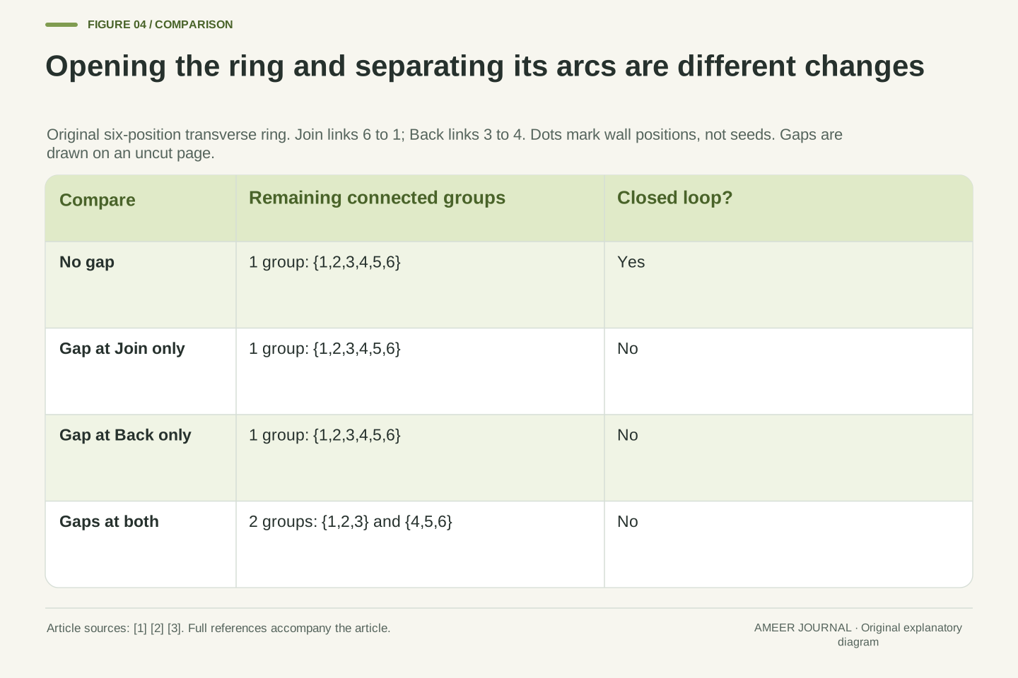
Start with both named connections present. You can travel around the entire drawn loop and return to your start. Next leave a gap only at Join. The loop is open, but you can still trace from dot one through two, three, four and five to six. All six dots remain in one connected wall path.
Leaving a gap only at Back gives the same count for a different reason: the remaining route passes through Join. The drawing is open but still connected. When both Join and Back are gaps, that last connection between the sides is gone. Dots one, two and three form one group; dots four, five and six form another.
The four cases therefore have one, one, one and two connected groups. An opening does not automatically mean the whole wall has become two separate pieces. Two suitably placed gaps in this simple ring do make two arcs. That is the precise relationship our model contributes to the explanation of two valve regions.
With no links removed, the graph is one connected cycle. Removing Join alone leaves the path 1–2–3–4–5–6, so the component count remains one. Removing Back alone leaves a different single path through Join. In both cases closedness changes while connected-component count does not.
Removing both named links leaves edge sets {1–2,2–3} and {4–5,5–6}. The components are therefore {1,2,3} and {4,5,6}. The four combinations have counts 1,1,1,2. These results follow from the declared adjacency list, not from an observed pod or a claim that the two seams must open simultaneously.
The equal three-marker groups arise because Join and Back were placed opposite one another in this six-position schematic. Their equal marker counts do not establish equal real areas, masses or seed distributions. The robust result is the distinction between opening a cycle at one location and separating its transverse wall path at two locations.
Turning a picture does not rename its anatomical regions
Imagine rotating the same drawing halfway around. Join may now appear at the bottom instead of the top. Back changes its page position too. Neither region has changed its identity: the label still follows the margin-joining region or the opposite back region that it named before.
This helps when two books show a pod from different directions. A line at the top of one picture need not be the same named line as the top of another. Read the labels and the viewpoint. Ventral and dorsal are anatomical terms in this carpel description [2][3], not instructions that every diagram must put a particular seam above the other.
Test your explanation by describing the picture without saying top, bottom, left or right. Try: Join is where the represented margins meet; Back is the other named region. Then trace the four opening cases again in the rotated view. The connections and group counts stay the same even though the drawing looks different on the page.
A rigid rotation of the schematic preserves adjacency and the identities attached to its labelled regions. Page orientation therefore cannot define ventral and dorsal. Their meanings refer to margin fusion and the dorsal region within the carpel model [2][3]. A change of drawing viewpoint must carry those anatomical labels with the represented structures.
The six-position ring also admits six cyclic relabellings. Applying the same relabelling to every surviving edge preserves each component count in all four opening states, giving 24 finite checks. This is an invariance of the defined graph. It is not a new anatomical measurement or a claim that real pods have six rotationally equivalent positions.
A useful diagram caption names its view and labels the regions directly. A reader should not have to infer structural identity from the direction a specimen happens to face in a photograph. This is especially helpful when distinguishing a longitudinal seam from its crossing point in a transverse schematic.
A cross-section cannot tell the whole story at the ends
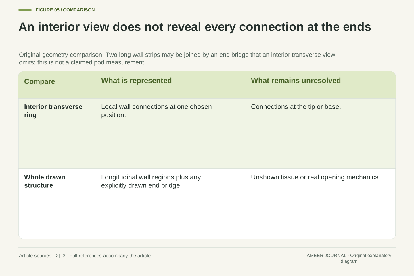
Our ring is a view across one interior place along the pod. It leaves out the tip and base. Two separate arcs in that view do not, by themselves, prove that the entire wall sections are disconnected everywhere. Something outside the chosen view could still join them. The model has no information about those missing locations.
Imagine two long drawn strips that are separate across their middles but joined by a bridge at one end. A view through the middle shows two pieces of outline. A view including the bridge shows how they remain connected elsewhere. This is another drawing example, not an extra claim about the exact attachments of a particular pea pod.
That boundary keeps our answer honest. We use the ring to explain the wall regions between two sutures and the change in their local connections. We do not use it to decide when a real valve falls away, how quickly a pod opens or which force moves it. A small diagram can answer a useful question without answering every question.
The graph models transverse connectivity away from the fruit ends. Two components in that section do not logically imply two disconnected three-dimensional objects. Additional connections outside the section can preserve global connectivity. Endpoints, attachments and the extent of each longitudinal opening are absent from the graph by definition.
A simple countermodel uses two longitudinal wall strips joined only at an end bridge. An interior transverse slice intersects two separate arcs, while the complete structure remains connected through the bridge. This construction establishes the limitation of the observation, not the actual end anatomy of a sampled pod.
Consequently, the source-supported two-valve terminology should not be turned into a guarantee of complete simultaneous detachment. The section model establishes an arrangement of wall regions and local opening states. Claims about global separation, time order or movement need additional anatomical or observational evidence rather than more confidence in the same incomplete view.
Other fruit plans show why one count is not enough
The university fruit guide contrasts legumes that open along two sutures with follicles that open along one [2]. An opening line is therefore a useful detail to describe. It is not a universal rule that every dry fruit must open in the same way or that each line stands for one separate original flower part.
Kew's comparison with another plant family, Bignoniaceae, describes commonly two-valved capsules associated with two fused carpels [4]. You do not need to memorize that family name. Its role here is a comparison: two opening wall sections can occur in a different anatomical plan from the one-carpel pea example.
Make a small table headed starting structure, opening description and wall sections. Put the pea example in one row and the specifically described comparison in another. Keep the labels supplied by the references instead of filling missing boxes from appearance. A shared number in the wall-section column does not force the starting-structure column to match.
The fruit-lab account distinguishes two-suture legume dehiscence from the ventral one-suture opening of follicles [2]. These descriptions concern opening pattern and must retain their fruit-type scope. They do not license inferring a carpel count by simply counting visible cracks in an unknown fruit.
The Kew-hosted Neotropikey comparison describes Bignoniaceae ovaries with two fused carpels and fruits usually forming dry capsules that dehisce by two valves [4]. Together with the monocarpellate legume, this provides a concrete reason that two valves alone cannot uniquely specify the developmental carpel count. The comparison remains qualified rather than universal for every family member.
A structural comparison should therefore retain separate fields for carpel organization, mature wall, opening pattern and observation scope. Inference proceeds from the full anatomical description, not one convenient number. This differs from merely sorting fruits as dry or fleshy: it asks which relationship produced the wall sections being counted.
A botanical pattern is not a promise about the next pod you see
Dehiscent means opening to release seeds; indehiscent means remaining closed in that sense [1]. These words describe mature fruit behavior. Our neat opening drawings explain a structural pattern. They do not predict that any green pod in a photograph will immediately open, or that every fruit called a pod follows the same sequence.
Kew's accounts record several alternatives within the legume group, including fruits that remain closed, open differently or divide into segments [4]. Those exceptions matter. The typical pea explanation can be useful without becoming a rule for every related plant. A common name or a similar outline is not enough to erase those differences.
If a picture does not show a seam clearly, write not shown instead of inventing an opening. If it shows a closed fruit, do not fill in a timetable for what happens next. A labeled anatomical drawing and a photograph of one moment provide different kinds of information. Our lesson uses the first to explain structure and leaves an actual pod's future unobserved.
Dehiscence describes mature fruit opening, while indehiscence describes retention within an unopened fruit wall [1]. The schematic does not model maturation or the material processes enabling separation. A structural two-suture account consequently does not yield an opening time, a force threshold or a guaranteed sequence for a particular immature fruit.
The Kew-hosted Flora of Tropical East Africa description of the papilionoid group states the usual two-valve condition alongside one-suture, indehiscent and transversely segmented alternatives [4]. The broader family account likewise records varied fruit types. Those qualifications are retained; historical subfamily classifications and numerical species totals are not imported as current taxonomy.
Distinguish absent evidence from evidence of absence. A seam not resolved in a photograph has not thereby been shown absent anatomically. Conversely, a conventional label in a teaching drawing does not establish that a particular specimen has opened along it. The required next information depends on whether the question concerns anatomical identity, present opening state or later behavior.
Finish with an explanation that follows the connections
Ask your partner to draw a single curved wall with Join and Back marked. Explain which region represents the meeting margins and which represents the opposite part of the same carpel. Then choose one of the four ring states and describe a surviving route between numbered dots. Pointing to the route makes the answer checkable.
Try the sentence: this drawing represents one carpel's wall, with two named opening regions. If both indicated connections are absent in the interior cross-section, two wall arcs remain. Add one limit: the drawing does not include the ends. You have now explained the structure, the change and the boundary of the picture.
Keep the page as a personal learning note. The next time a diagram shows two visible parts, the useful question is what kind of parts they are and how they are connected. For a pea pod, two sutures and two valve regions can belong to the fruit of one carpel. The labels and the traced relationships explain why.
A complete explanation can name the anatomical premise, the representation and the derived result separately. The premise is the source-supported monocarpellate pea legume [1][2]. The representation identifies fused margins as Join and the dorsal region as Back. The derivation shows that removing both named transverse links leaves two wall arcs in the defined ring.
Check the countercases as well: with neither link absent the ring is closed and connected; with either single link absent it is open but connected; with both absent it has two components. These distinctions explain why a visible opening, a valve count and a carpel count cannot be treated as interchangeable observations.
Finally retain the scope: arbitrary drawing coordinates, interior cross-section, no end attachments, no opening dynamics and no specimen identification from shape alone. The useful learning outcome is a traceable relation between one floral unit and two different opening regions. It is stronger than memorizing a count because it explains which change in the drawing produces each result.
Sources & further reading
Explore the references behind this guide. External pages may change after our review.
- 8.1 Fruit Morphology – The Science of PlantsUniversity of Minnesota · Accessed 7 Nov 2026
- Plant Taxonomy - Biology 308Stephen G. Saupe / College of Saint Benedict and Saint John’s University · Accessed 7 Nov 2026
- Plant Taxonomy - Biology 308Stephen G. Saupe / College of Saint Benedict and Saint John’s University · Accessed 7 Nov 2026
- Fabaceae Lindl. | Plants of the World Online | Kew ScienceRoyal Botanic Gardens, Kew · Accessed 7 Nov 2026
Article updated 7 Nov 2026.